Introduction to Photoelectric Sensors in ECG Machines
Photoelectric sensors play a pivotal role in the functionality and accuracy of modern electrocardiograph (ECG) machines. These sensors convert light signals into electrical signals, enabling precise detection of physiological changes. In ECG applications, they are primarily used in optical heart rate monitoring, lead detection, and signal isolation. This integration enhances patient safety and data reliability, addressing critical requirements in clinical and emergency settings.
Operating Principle and Key Components
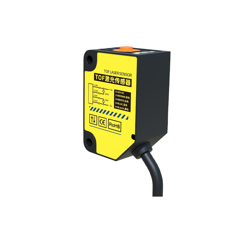
The core principle relies on photoelectric effect, where light-emitting diodes (LEDs) and photodetectors work together. In an ECG system, photoelectric sensors often employ reflective or transmissive modes to detect heart rate via tissue perfusion. Key components include a high-sensitivity photodiode, a modulated light source, and a transimpedance amplifier. The sensor operates by emitting a specific wavelength, typically green or infrared, and analyzing the backscattered light changes due to blood volume variations.
Advantages Over Traditional Electrode-Based Systems
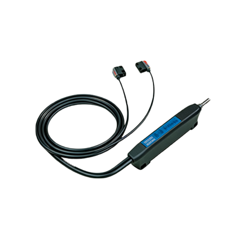
Compared to conventional wet electrode systems, photoelectric sensors offer non-invasive, motion-tolerant, and long-term monitoring capabilities. They eliminate skin irritation and signal degradation caused by electrode gel drying out. In ECG machines, these sensors enable continuous monitoring during patient movement, which is crucial for stress tests and ambulatory ECGs. Additionally, optical isolation reduces electrical interference, improving signal-to-noise ratio in high-voltage environments like defibrillators.
Integration with ECG Signal Processing
Photoelectric sensors are often integrated into the front-end circuit of ECG machines, providing galvanic isolation between the patient and the measurement system. This isolation is achieved through optocouplers, which use phototransistors to transmit heart rate data while preventing ground loops. The sensor output is then processed by a microcontroller using algorithms for motion artifact rejection and peak detection, ensuring accurate R-wave identification in arrhythmia analysis.
Design Considerations for Clinical Use
For reliable performance in clinical settings, photoelectric sensors must meet stringent standards for sensitivity, response time, and ambient light rejection. Engineers design these sensors with narrow-band optical filters and synchronous demodulation to minimize interference from room lighting. Thermal management is also critical, as prolonged use may cause LED heating affecting sensor drift. Housing materials should be medical-grade, biocompatible, and easy to clean.
Future Trends and Innovations
Emerging trends include multi-wavelength photoelectric sensors for better tissue penetration and multi-lead ECG integration. Advances in microelectromechanical systems (MEMS) allow miniaturized sensors for wearable ECG patches. Furthermore, machine learning algorithms are being developed to enhance the extraction of cardiac features from photoplethysmogram signals. These innovations promise to make photoelectric ECG systems more accessible and accurate for remote patient monitoring.
Conclusion
Photoelectric sensors are indispensable in advancing ECG technology, providing robust performance, safety, and patient comfort. As the medical industry pushes toward non-invasive and continuous monitoring, these sensors will continue to evolve, driving improvements in diagnostic accuracy and user experience. For electrical engineers, understanding these components is essential for designing next-generation cardiac care devices.